Tiedosto:Staphylococcus aureus VISA 2.jpg

Alkuperäinen tiedosto (1 420 × 1 091 kuvapistettä, 259 KiB, MIME-tyyppi: image/jpeg)
| Kuvaus |
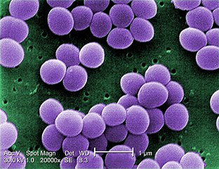
English: Under a very high magnification of 20,000x, this scanning electron micrograph (SEM) shows a strain of Staphylococcus aureus bacteria taken from a vancomycin intermediate resistant culture (VISA). Under SEM, one can not tell the difference between bacteria that are susceptible, or multidrug resistant, but with transmission electron microscopy (TEM), VISA isolates exhibit a thickening in the cell wall that may attribute to their reduced susceptibility to vancomycin . See PHIL 11156 for a black and white version of this image. VISA and VRSA are specific types of antimicrobial-resistant staph bacteria. While most staph bacteria are susceptible to the antimicrobial agent vancomycin some have developed resistance. VISA and VRSA cannot be successfully treated with vancomycin because these organisms are no longer susceptibile to vancomycin. However, to date, all VISA and VRSA isolates have been susceptible to other Food and Drug Administration (FDA) approved drugs. How do VISA and VRSA get their names? Staph bacteria are classified as VISA or VRSA based on laboratory tests. Laboratories perform tests to determine if staph bacteria are resistant to antimicrobial agents that might be used for treatment of infections. For vancomycin and other antimicrobial agents, laboratories determine how much of the agent it requires to inhibit the growth of the organism in a test tube. The result of the test is usually expressed as a minimum inhibitory concentration (MIC) or the minimum amount of antimicrobial agent that inhibits bacterial growth in the test tube. Therefore, staph bacteria are classified as VISA if the MIC for vancomycin is 4-8µg/ml, and classified as VRSA if the vancomycin MIC is >16µg/ml. |
||
| Päiväys | |||
| Lähde |
|
||
| Tekijä |
Content Providers(s): CDC/ Matthew J. Arduino, DRPH |
||
| Käyttöoikeus (Tämän tiedoston uudelleenkäyttö) |
PD-USGov-HHS-CDC English: None - This image is in the public domain and thus free of any copyright restrictions. As a matter of courtesy we request that the content provider be credited and notified in any public or private usage of this image. |
Tämän teoksen on valmistanut Yhdysvaltain tartuntatautien valvonta- ja ehkäisykeskusten (Centers for Disease Control and Prevention, CDC) työntekijä osana kyseisen työntekijän virkatointa. Yhdysvaltain liittovaltion viranomaisten työntekijöiden tekemät teokset eivät saa tekijänoikeuden suojaa Yhdysvaltain tekijänoikeuslain 105 § mukaisesti.
|
Tiedoston historia
Päiväystä napsauttamalla näet, millainen tiedosto oli kyseisellä hetkellä.
| Päiväys | Pienoiskuva | Koko | Käyttäjä | Kommentti | |
|---|---|---|---|---|---|
| nykyinen | 4. elokuuta 2009 kello 02.24 | | 1 420 × 1 091 (259 KiB) | Raeky | {{Information |Description={{en|1='''Under a very high magnification of 20,000x, this scanning electron micrograph (SEM) shows a strain of Staphylococcus aureus bacteria taken from a vancomycin intermediate resistant culture (VISA).'''<p> Under SEM, one |
Tiedoston käyttö
Seuraava sivu käyttää tätä tiedostoa:
Tiedoston järjestelmänlaajuinen käyttö
Seuraavat muut wikit käyttävät tätä tiedostoa:
- Käyttö kohteessa ar.wikipedia.org
- فيلقية
- أشعار بكتيرية
- آزوتية
- ريكتسيا
- مستجذرة
- مفطورة
- ملوية (جنس)
- وتدية خناقية
- متفطرة
- مطثية حاطمة
- هدب (بكتيريا)
- شعاوات
- متفطرة جذامية
- مرق السيلينيت
- بوريليا برغدورفيرية
- مكورات
- صبغة أورامين رومدامين
- اختبار الأكسيداز
- انزلاق بكتيري
- ملتويات (بكتيريا)
- ملتوية معوية
- نستق
- قالب:بذرة بكتيريا
- نوستك
- مستدمية
- كليبسيلا
- سفينغوبيوم
- جرثومة مخاطية
- متقلبات
- متقلبات زيتا
- متقلبات إيبسيلونية
- متقلبات غاما
- متقلبات بيتا
- متقلبات ألفا
- بكتيريا مغزلية
- عصوانيات
- خضربيات
- دليل برجاي لعلم الجراثيم المنهجي
- جراثيم ثؤلولية
- مستعلقات
- سحناوات
- متمصرة
- بكتيريا حمضية
- متدثرات
- كلورو بكتيريا
- شبكيات الكبب
- شبكية الكبة حرارية
- ليفيات
- سلسلية
- سلسلية طوقية الشكل
Näytä lisää tämän tiedoston järjestelmänlaajuista käyttöä.